AI Breakthroughs in Healthcare: August 2026 — From Liver Diagnosis to Non-Invasive ICU Monitoring
August 2026 will be remembered as one of the most consequential months in the history of AI in healthcare. Within a single week, five landmark studies appeared — spanning liver malignancy diagnosis, retinal disease classification, non-invasive ICU monitoring, heart obstruction detection, and large language model clinical decision support. Together, they demonstrate that AI is no longer confined to retrospective benchmarks; it is operating in prospective clinical trials, real-world hospital workflows, and wearable monitoring systems.
For researchers working in computer vision and deep learning — as I have throughout my doctoral work on pedestrian detection — these advances are directly relevant. Medical imaging AI relies on the same foundations: object detection, semantic segmentation, feature extraction, and classification. The architectural innovations that power pedestrian detectors also power liver and retinal diagnosis. And the shift from detection to diagnosis in healthcare mirrors the evolution in autonomous driving — from identifying objects to understanding scenes.
In this article, I review each of the five breakthroughs in depth, explain their core technical contributions, and discuss how they connect to broader themes in computer vision research.
| Value | Metric |
|---|---|
| 0.975 | LiON AUC multicenter validation |
| 10,333 | Patients in LiON prospective trial |
| 97.46% | FOCUS F1 score abnormality detection |
| 51 | Overlooked lesions caught by AI (LiON) |
| 28 | ICU patients in MOSAIC study |
| 99% | SHAKED outputs clinically appropriate |
1. LiON: AI-Guided Liver Malignancy Diagnosis at Scale
On August 19, 2026, Nature Medicine published a study that sets a new benchmark for AI-guided cancer diagnosis. The paper, “Large-scale AI-guided liver malignancy diagnosis: multicenter study and a single-arm trial,” introduces LiON, an AI system trained to identify malignant liver lesions on imaging scans. The scale and rigor of the evaluation are unprecedented.
Training and Retrospective Validation
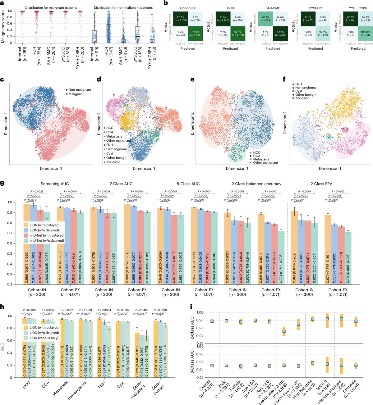
LiON was trained on 6,443 patients and then validated retrospectively across 22,251 patients spanning multiple centers and real-world cohorts. In multicenter validation, LiON achieved an AUC of 0.975 (95% CI: 0.971–0.979) — a figure that places it among the highest-performing diagnostic AI systems ever reported in any medical imaging domain.
Crucially, the system maintained strong performance even in patients with conditions that typically degrade imaging quality and diagnostic confidence. In patients with hepatic steatosis (fatty liver disease), LiON achieved an AUC of 0.971. In patients with cirrhosis — one of the most challenging populations for liver imaging due to nodular distortion and altered vascularity — it still achieved an AUC of 0.924.
Prospective Single-Arm Trial
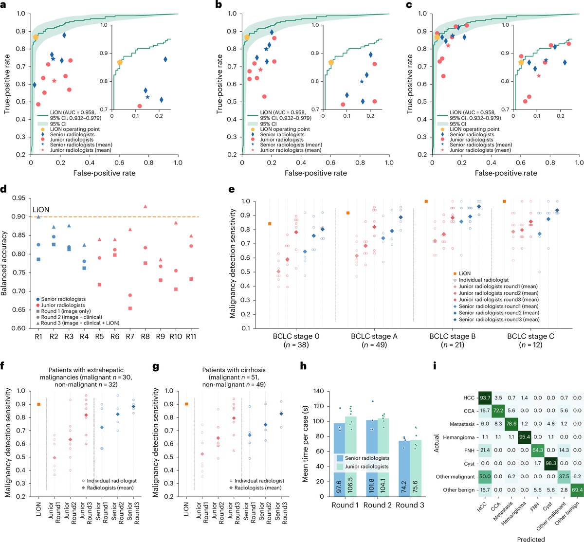
The retrospective results are impressive, but what sets LiON apart is the prospective single-arm trial involving 10,333 patients in routine clinical practice. This is not a benchmark evaluation on curated datasets — it is a real-world deployment in actual hospital workflows. The prospective trial AUC was 0.952 (95% CI: 0.942–0.961), a modest but expected drop from the retrospective AUC, confirming that the system generalizes to the noise, variability, and edge cases of clinical reality.
AI-Human Collaboration: Beyond Accuracy Metrics
Perhaps the most striking finding is what happened when LiON was paired with radiologists. In the prospective trial, AI-human collaboration identified 51 previously overlooked lesions, of which 15 were malignancies. These were cancers that the standard clinical workflow had missed. The AI detections triggered 37 amended radiology reports, 22 multidisciplinary team escalations, and direct changes in clinical management for affected patients.
Key Results from LiON
Trained on 6,443 patients and validated prospectively on 10,333, LiON achieved AUC 0.975 in multicenter validation and AUC 0.952 in a prospective clinical trial. AI-human collaboration caught 15 previously missed malignancies, triggering 37 amended reports and 22 multidisciplinary team escalations. The system maintained strong performance even in patients with hepatic steatosis (AUC 0.971) and cirrhosis (AUC 0.924). Read the paper (Nature Medicine) →
Connection to Computer Vision
LiON is fundamentally a medical object detection and classification system — the same class of problem that pedestrian detection addresses. The lesion detection stage identifies suspicious regions in the liver scan (analogous to detecting pedestrians in a traffic scene), and the classification stage determines whether the detected region is malignant (analogous to classifying a detected object as pedestrian vs. non-pedestrian). The architectural choices that make LiON robust to steatosis and cirrhosis — conditions that alter the visual appearance of the organ — are directly parallel to the challenge of making pedestrian detectors robust to adverse weather, where fog, rain, and low light similarly alter the visual characteristics of the scene.
| System | Target | Study Size | Key Metric | Published |
|---|---|---|---|---|
| LiON | Liver malignancy | 22,251 + 10,333 | AUC 0.975 | Nature Medicine |
| FOCUS | Retinal disease | 3,300 + 1,345 | F1 94.39% | npj Digital Med |
| MOSAIC | ICU blood pressure | 28 | Matched invasive | Comp Bio Med |
| Mayo AI | Heart obstruction | 1,833 + 275 + 46 | Outperformed experts | Circulation |
| SHAKED | ED decision support | 1,138 | 99% appropriate | Nature Medicine |
2. FOCUS: Foundation Model-Driven Retinal Disease Diagnosis
Also published on August 19, 2026, in npj Digital Medicine, the FOCUS system (Full-process OCT-based Clinical Utility System) represents a different paradigm: instead of a narrow, task-specific detector, FOCUS is a foundation model that automates the entire diagnostic workflow for 3D optical coherence tomography (OCT) retinal scans.
End-to-End Workflow Automation
Unlike most medical AI systems that perform a single task — detect a disease, classify a scan, measure a parameter — FOCUS handles the full diagnostic pipeline:
- Quality assessment: Is the scan of sufficient quality to be diagnostic? (F1: 99.01%)
- Abnormality detection: Does the scan show any signs of disease? (F1: 97.46%)
- Patient-level diagnosis: What specific disease(s) are present? (F1: 94.39%)
This three-stage pipeline mirrors how an ophthalmologist actually reads a scan: first assessing quality, then scanning for abnormalities, then arriving at a specific diagnosis. By automating all three stages, FOCUS moves beyond “AI as a second reader” toward “AI as a complete diagnostic pathway.”
External Validation Across Devices and Centers
FOCUS was trained on 3,300 patients (40,672 slices) and externally validated on 1,345 patients (18,498 slices) across four centers using diverse OCT devices. In real-world external validation, FOCUS achieved an F1 range of 90.22% to 95.24% — remarkably consistent across different hardware, patient populations, and clinical settings.
Most notably, FOCUS matched expert performance. On abnormality detection, FOCUS scored 95.47% F1 compared to experts at 90.91%. On multi-disease diagnosis, FOCUS scored 93.49% F1 compared to experts at 91.35%. In both cases, the AI not only reached human-level performance but slightly exceeded it — a result that, if replicated across ophthalmology, could fundamentally change how retinal diseases are screened and diagnosed.
Key Results from FOCUS
FOCUS automates the full retinal diagnostic workflow: quality assessment (F1 99.01%), abnormality detection (F1 97.46%), and patient-level diagnosis (F1 94.39%). Externally validated on 1,345 patients across four centers and diverse OCT devices, it matched or exceeded expert ophthalmologists: abnormality detection 95.47% vs. 90.91% and multi-disease diagnosis 93.49% vs. 91.35%. Read the paper (npj Digital Medicine) →
Connection to Computer Vision
FOCUS is a foundation model — a single, large, pre-trained model that handles multiple tasks rather than requiring separate models for each. This is the same paradigm shift transforming general-purpose computer vision. Models like CLIP, DINO, and SAM have shown that a single well-trained vision backbone can be adapted to detection, segmentation, and classification with minimal fine-tuning. FOCUS demonstrates that this paradigm works in medical imaging as well, and the implications are profound: instead of training dozens of narrow models for each retinal disease, a single foundation model can handle the entire diagnostic pathway. For computer vision research, FOCUS validates the foundation model approach in a domain where the cost of error is measured in vision loss rather than missed frames.
3. MOSAIC: Non-Invasive Blood Pressure Monitoring with Wearable Sensors
On August 17, 2026, researchers at Johns Hopkins University published a study in Computers in Biology and Medicine that could change how blood pressure is monitored in intensive care units worldwide. The system, called MOSAIC, uses two simple wearable sensors and a deep learning model to replace one of the most invasive and risk-laden procedures in critical care.
The Problem with Arterial Lines
In ICU settings, continuous blood pressure monitoring typically requires an intra-arterial catheter — a thin tube inserted directly into an artery. While this provides accurate, beat-by-beat blood pressure readings, it carries significant risks:
- Bleeding: Arterial catheters can cause hematoma formation and hemorrhage
- Clotting: Thrombus formation around the catheter can obstruct blood flow
- Infection: Any invasive line is a potential entry point for bloodstream infections
- Pain and discomfort: Arterial cannulation is painful and limits patient mobility
How MOSAIC Works
MOSAIC replaces the arterial line with two non-invasive wearable sensors: a chest ECG sensor that captures the electrical activity of the heart, and a finger photoplethysmography (PPG) sensor that measures blood volume changes in the peripheral vasculature using light. A deep learning model then converts these two signals into a continuous arterial blood pressure waveform — the same type of output that an invasive arterial catheter provides.
The validation was conducted in 28 ICU patients at Johns Hopkins Hospital, and the results showed that the MOSAIC waveforms closely matched the invasive arterial catheter readings. The deep learning model learned the complex, nonlinear relationship between the ECG and PPG input signals and the true arterial blood pressure — a physiological mapping that would be nearly impossible to express with traditional signal processing rules.
Beyond the ICU
The researchers note that MOSAIC’s potential extends well beyond the ICU. Because the sensors are small, inexpensive, and non-invasive, the system could enable:
- Continuous monitoring on general hospital wards where arterial lines are impractical
- At-home hypertension monitoring that provides the same waveform fidelity as an arterial line, without the risks
- Ambulatory blood pressure tracking for chronic disease management, capturing the full hemodynamic picture rather than isolated cuff readings
Key Results from MOSAIC
MOSAIC uses two wearable sensors (chest ECG + finger PPG) and a deep learning model to generate continuous arterial blood pressure waveforms that closely match invasive arterial catheter readings. Validated in 28 ICU patients at Johns Hopkins Hospital, the system could eliminate the need for arterial lines and their associated risks of bleeding, clotting, and infection, with potential applications in general ward monitoring and at-home hypertension tracking. Read the study (Johns Hopkins) →
Connection to Computer Vision
While MOSAIC is not a vision system, it exemplifies a broader principle that is central to deep learning research: the ability of neural networks to learn complex, nonlinear mappings from raw sensor signals to clinically meaningful outputs. The same deep learning architectures — convolutional and recurrent layers, attention mechanisms, and adaptive feature extraction — that enable MOSAIC to map ECG+PPG signals to blood pressure waveforms also enable pedestrian detectors to map RGB pixels to bounding boxes. The underlying principle is the same: learn the mapping from raw data to the desired output, end-to-end, without hand-crafted intermediate representations.
4. Mayo Clinic: AI Detection of Heart Obstruction from Routine Ultrasound
On August 19, 2026, Mayo Clinic published a study in Circulation: Cardiovascular Imaging that removes one of the key barriers to diagnosing a life-threatening heart condition: the need for specialized Doppler expertise.
The Clinical Challenge
Hypertrophic cardiomyopathy (HCM) is a condition in which the heart muscle becomes abnormally thick, potentially causing left ventricular outflow tract (LVOT) obstruction — a blockage that restricts blood flow from the heart. Detecting LVOT obstruction traditionally requires Doppler echocardiography, a specialized ultrasound technique that measures blood flow velocity. Doppler interpretation requires significant expertise, and this expertise is not available in many clinical settings, particularly in resource-limited environments and point-of-care scenarios using portable ultrasound.
The AI Solution
The Mayo Clinic team developed an AI model that identifies LVOT obstruction using only routine non-Doppler B-mode ultrasound videos — the standard 2D grayscale ultrasound images that any echocardiography machine can acquire, without requiring Doppler expertise. The model was trained and tested on a large dataset:
- Training: 1,833 patients at Mayo Clinic
- Internal test: 275 patients
- External validation: 46 patients from a South Korean hospital — a different country, different patient population, and different ultrasound equipment
In some cases, the AI outperformed two expert echocardiographers reviewing the same non-Doppler images. This result is remarkable: it suggests that the model is extracting visual information from the B-mode video that even trained human eyes miss when Doppler data is not available.
Expanding Access to Screening
The clinical implications are significant. Because B-mode ultrasound is the most widely available cardiac imaging modality — available even on portable point-of-care ultrasound devices — the AI model could enable LVOT obstruction screening in settings where Doppler expertise is unavailable. This includes:
- Rural hospitals and clinics in low-resource settings
- Emergency departments using handheld ultrasound
- Primary care offices performing screening echocardiograms
- Sports medicine screenings for athletes at risk of sudden cardiac death
Key Results from Mayo Clinic AI
The AI model identifies LVOT obstruction in HCM patients using only routine B-mode ultrasound — no specialized Doppler needed. Trained on 1,833 patients, tested on 275, and externally validated on 46 patients from a South Korean hospital. In some cases, the AI outperformed two expert echocardiographers reviewing the same non-Doppler images, potentially expanding screening to settings where Doppler expertise is unavailable, including portable ultrasound devices. Read the study (Mayo Clinic) →
Connection to Computer Vision
This study is a pure computer vision problem solved with deep learning. The model processes 2D video sequences (ultrasound frames) and learns to identify a dynamic anatomical phenomenon (LVOT obstruction) from spatio-temporal patterns. This is structurally identical to video-based object detection and action recognition in computer vision — the same class of problem that appears in traffic scene analysis, where the system must detect and classify dynamic events from a video stream. The fact that the AI outperforms experts on the same visual input demonstrates that deep feature extraction can surpass human visual perception even in a domain where human experts have years of training.
5. SHAKED: When Accuracy Is Not Enough — The Human Side of AI in Emergency Medicine
The fifth study, also published on August 19, 2026 in Nature Medicine, is perhaps the most important for understanding what it actually takes to deploy AI in clinical practice. The paper, “Prospective evaluation of a large language model clinical decision support system in the emergency department,” introduces SHAKED and reveals a finding that should temper every hype cycle about AI in healthcare.
The Study Design
SHAKED was evaluated prospectively at Rambam Health Care Campus in Israel across 1,138 patients over 4 weeks, running in two parallel emergency department units. This is a rigorous real-world evaluation — not a retrospective benchmark, not a simulation, but an actual deployment in a working emergency department.
The Technical Results: Excellent
From a technical standpoint, SHAKED performed well. 99 out of 100 sampled outputs were rated clinically appropriate. Physicians showed a clear preference for SHAKED when seeking radiology consultations (odds ratio = 2.98). And there was a non-significant trend toward shorter consultation cycle time (−9.4 minutes, P = 0.077).
The Adoption Results: Alarming
Despite the excellent technical performance, clinical adoption declined from 68% to 30% over the four-week study period. Physicians simply stopped using the system. The decline was not driven by inaccuracy or safety concerns — no adverse events were detected. Instead, it was driven by workload-sensitive disengagement: when the emergency department got busier, physicians had less cognitive bandwidth to engage with the AI system, even when it was helpful.
This finding reframes the entire conversation about AI in healthcare. The field has focused overwhelmingly on algorithmic accuracy — AUC scores, F1 metrics, sensitivity and specificity. SHAKED demonstrates that sustained clinician engagement, not algorithmic accuracy, is the key barrier. You can build the most accurate AI system in the world, and it will still fail to improve outcomes if clinicians do not use it.
Key Results from SHAKED
In a prospective evaluation across 1,138 emergency department patients, SHAKED achieved 99/100 clinically appropriate outputs and a strong physician preference for radiology consultations (OR = 2.98). But clinical adoption declined from 68% to 30% over just four weeks — driven not by inaccuracy but by workload-sensitive disengagement. The key insight: sustained clinician engagement, not algorithmic accuracy, is the key barrier to AI adoption in healthcare. Read the paper (Nature Medicine) →
Connection to Computer Vision and Autonomous Systems
The SHAKED result has direct implications for autonomous driving and safety systems. In both healthcare and autonomous driving, the human operator is the final decision-maker, and the AI system is a support tool. If emergency physicians — highly trained professionals working in a structured environment — disengage from an AI system when workload increases, what happens to a driver in a stressful traffic situation? The lesson is clear: building an accurate system is necessary but insufficient. The system must also be designed for sustained engagement under real-world workload conditions — low cognitive overhead, minimal interaction cost, and seamless integration into existing workflows.
Synthesizing the Breakthroughs: Common Themes and Broader Implications
Individually, each of these studies represents a significant advance. Together, they reveal several important themes:
1. The Shift from Benchmarks to Prospective Clinical Trials
Three of the five studies (LiON, FOCUS, SHAKED) include prospective clinical evaluations — real patients, real clinical workflows, real-world conditions. This represents a maturation of the field from retrospective benchmark-chasing to genuine clinical validation. LiON’s 10,333-patient prospective trial and FOCUS’s four-center external validation are particularly notable for their scale and rigor.
2. Foundation Models Are Transforming Medical Imaging
FOCUS demonstrates that the foundation model paradigm — a single pre-trained model that handles multiple related tasks — works in medical imaging just as it does in general computer vision. This is significant because the medical AI field has traditionally relied on narrow, task-specific models that require separate training data and validation for each disease. A foundation model that can handle the entire diagnostic workflow, from quality control to multi-disease classification, is far more deployable than a collection of single-task detectors.
3. AI-Human Collaboration Beats AI Alone
LiON’s result — that AI-human collaboration caught 51 previously missed lesions, including 15 malignancies — underscores that the most impactful deployment model is not AI replacing clinicians, but AI augmenting clinicians. The 37 amended reports and 22 multidisciplinary team escalations are concrete patient outcomes, not abstract metrics.
4. Deployment Barriers Are Human, Not Technical
SHAKED’s finding that clinical adoption halved in four weeks despite near-perfect accuracy is the most important cautionary result of the month. It applies not only to healthcare but to any domain where AI assists human decision-making under time pressure. The deployment problem is not “can the AI be accurate?” but “will the human actually use it?”
5. Non-Invasive Is the Future
Both MOSAIC (wearable blood pressure) and the Mayo Clinic study (B-mode ultrasound instead of Doppler) demonstrate that AI can extract more information from less invasive sensors. This is a fundamental shift: instead of requiring more specialized and invasive data, AI makes existing, widely available data more informative. The parallel to computer vision is direct — just as MOSAIC extracts arterial blood pressure from simple ECG and PPG signals, modern pedestrian detectors extract rich scene understanding from simple RGB camera feeds, without requiring LiDAR or specialized sensors.
What These Breakthroughs Mean for Computer Vision Research
For the computer vision research community, and for my own work on pedestrian detection, these healthcare breakthroughs offer several concrete lessons:
- Foundation models are the future. Just as FOCUS shows that a single model can handle the full retinal diagnostic workflow, the future of pedestrian detection lies not in training narrow detectors for specific conditions (fog, rain, night) but in foundation models that adapt to diverse conditions with minimal fine-tuning.
- Prospective validation is non-negotiable. LiON’s 10,333-patient prospective trial and SHAKED’s real-world deployment study demonstrate that retrospective benchmarks are necessary but insufficient. For pedestrian detection, this means testing systems not just on curated datasets but in real driving conditions, with real-time constraints and real sensor noise.
- Human engagement determines impact. SHAKED’s adoption decline is a warning for any AI system designed to assist human operators. For pedestrian detection in autonomous driving, the lesson is that alert fatigue and cognitive overload are real risks. If a system generates too many false alarms, drivers will disengage — just as SHAKED’s physicians did.
- Less invasive, more informative. The trend toward extracting more from less — MOSAIC from two wearable sensors, Mayo Clinic AI from B-mode ultrasound — parallels the goal of building lighter, more efficient pedestrian detectors that run on edge devices without requiring expensive sensor arrays.
Looking Ahead: The Convergence of Medical and General-Purpose Vision
August 2026 marks a clear inflection point. The same deep learning architectures — convolutional networks, vision transformers, attention mechanisms, adaptive feature pyramids — that power pedestrian detection are now powering liver diagnosis, retinal screening, blood pressure monitoring, and heart obstruction detection. The foundation model paradigm that is transforming general computer vision is now transforming medical imaging as well.
But the most important lesson from this month is not about architecture — it is about deployment. LiON, FOCUS, MOSAIC, and the Mayo Clinic model all demonstrate that the technology works. SHAKED reminds us that technology that is not used does not help anyone. As we build ever more accurate AI systems, in healthcare and in AI research in Morocco and beyond, we must invest equally in understanding how these systems are adopted, sustained, and integrated into the workflows of the people they are designed to assist.
The future of AI in healthcare — and in every safety-critical domain — depends not just on what the algorithm can do, but on whether the human beside it will keep using it when it matters most.
Collaborate on AI & Healthcare Research
Interested in the intersection of computer vision, deep learning, and medical imaging? Dr. Razzok welcomes research partnerships, academic supervision inquiries, and joint publications.
Get in Touch →